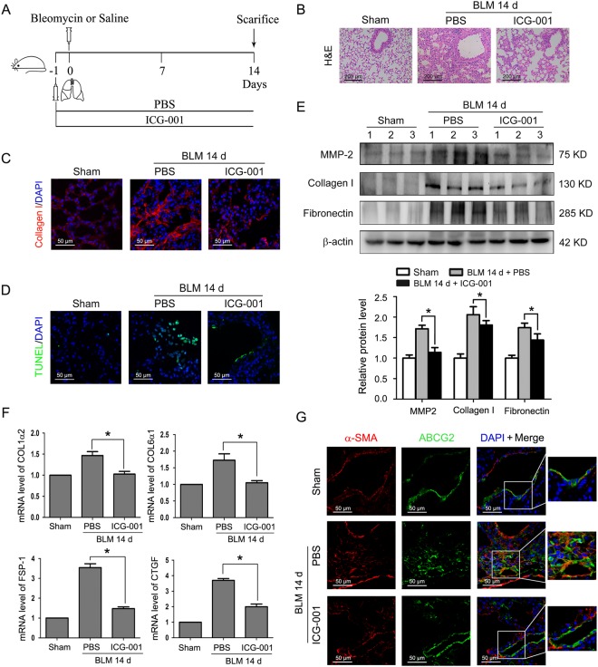
Figure 6.
ICG-001 attenuates pulmonary fibrosis and myofibroblast differentiation of LR-MSCs in a bleomycin-induced mouse fibrosis model. (A) Schematic presentation of the preventive study design. Mice (n = 6 in each group) started receiving daily intraperitoneal injection of PBS or ICG-001 (5 mg/kg body weight) one day before intratracheal instillation of saline or bleomycin (BLM) (5 mg/kg body weight). Mice were sacrificed on day 14. (B) The lung sections from the mice that have undergone different treatments were stained by hematoxylin-eosin (H&E) (C) The expression of collagen I in lungs was examined by immunofluorescence assay. Nuclei were stained with DAPI (blue). (D) Apoptotic cells of lung tissues were determined by terminal deoxynucleotidyl transferase-mediated dUTP nick end labeling (TUNEL) assay. Nuclei were stained with DAPI (blue) and green dots indicate apoptotic cells. (E) The protein levels of matrix metalloproteinase-2 (MMP-2), collagen I and fibronectin in lungs were determined by Western blot. Representative gel electrophoresis bands are shown, and the expression levels of proteins were quantified by densitometry and normalized to the expression of β-actin. Densitometry data are shown as mean ± SD. *P < 0.05 versus BLM 14 d + PBS. (F) Changes of the COL1α2, COL6α1, fibroblast-specific protein1 (FSP-1) and connective tissue growth factor (CTGF) transcripts in lungs were measured by Q-PCR. Results are shown as mean ± SD. *P < 0.05 versus BLM 14 d + PBS. (G) Myofibroblast differentiation of mouse LR-MSCs in vivo was performed using dual immunofluorescence staining to detect ATP-binding cassette transporter subtype G 2 (ABCG2) (green) and α-SMA (red). Nuclei were stained with DAPI (blue).

