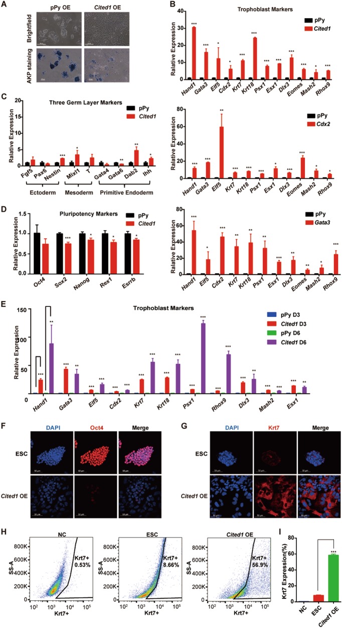
Fig. 3. Ectopic Cited1 activates trophoblast lineage genes.
a Typical morphological changes and AKP staining in E14T ESCs after overexpressing Cited1 for 2 days. Scale bar: 100 μm. b Results of qRT-PCR analysis of expression levels of trophoblast markers in ESCs overexpressing Cited1, Cdx2, or Gata3 for 3 days. The average mRNA level in cells transfected with the control vector pPy was set at 1.0. Data are shown as mean ± SD (n = 3). *p < 0.05, **p < 0.01, ***p < 0.001. c, d Results of qRT-PCR analysis of expression levels of three germ layer markers (c) and pluripotency-associated markers (d) in ESCs overexpressing Cited1 for 3 days. The average mRNA level in cells transfected with the control vector pPy was set at 1.0. Data are shown as mean ± SD (n = 3). *p < 0.05, **p < 0.01, ***p < 0.001. e qRT-PCR analysis of expression levels of trophoblast markers after transfection of plasmids as indicated in ESCs over a time course. The average mRNA level in cells transfected with the control vector pPy was set at 1.0. Data are shown as mean ± SD (n = 3). **p < 0.01, ***p < 0.001. f, g Immunofluorescence staining of ESCs after transfection of Cited1 for 6 days. Samples were stained with anti-Oct4 antibody (red) (e) and anti-Krt7 antibody (red) (f), respectively. DAPI staining highlights the nuclei (blue). Scale bar: 50 μm. h Flow cytometry density plots for Krt7 expression in ESCs after transfection of Cited1 for 6 days. Cells were fully dispersed, fixed, and immunostained for Krt7. For the negative control (NC), cells were exposed only to secondary antibody without prior exposure to the primary Krt7 antibody. i The statistical analysis of flow cytometry data for percentages of cells positive for Krt7 expression in ESCs overexpressing Cited1 or an empty vector for 6 days. Data are shown as mean ± SD (n = 3). ***p < 0.001

