Figure 5.

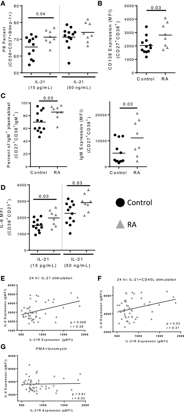
IL-21 mediated plasmablast differentiation is enhanced in RA. Naïve B cells were isolated from PBMC of control (n = 12) and RA patients (n = 8) and cultured with IL-21 (15 pg/mL or 50 ng/mL) and CD40L for 12 days. (A) Plasmablast percent was measured by staining for CD38+CD27+Blimp-1+ cells following 12 days of culture with either low (15 pg/ml) or high (50 ng/ml) levels of IL-21 in addition to CD40L. (B) CD138 expression was determined on plasmablasts (CD38+CD27+) from RA subjects and controls following high dose IL-21 and CD40L culture. (C) Frequency of IgM+ plasmablasts (CD38+CD27+) were assessed following 12 days of culture with high dose IL-21 and CD40L (left). IgM gMFI was assessed in plasmablasts by intracellular staining (right). (D) IL-6 gMFI was assessed on plasmablasts in response to low or high dose IL-21 in addition to CD40L stimulation in controls and RA subjects. (E) IL-21 expression correlates with IL-6 production following IL-21 stimulation (F) and IL-21+CD40L stimulation but not (G) PMA+Ionomycin stimulation. PBMCs from RA subjects (n = 52) were stimulated with IL-21 (E) or IL-21+CD40L (F) for 24 h. In the last 4 h of incubation, brefeldin A and monensin were added to prevent export of IL-6. The level of IL-6 was measured by flow cytometry on total B cells. IL-6 production was measured by flow cytometry. IL-21R expression was measured at day 0 on memory B cells. Black circles are controls and gray triangles are RA patients. Significance was assessed using Mann Whitney U tests. Correlation was assessed with the Pearson correlation.
