Figure 4.
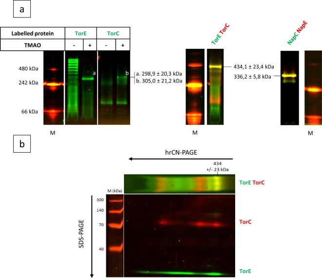
In vivo identification of the TorE-TorC complex. (a) hrCN-gels, first dimension. The strains were growth in anaerobiosis. The fluorescent labelled proteins were solubilized from the membrane extracts of the various recombinant strains using DDM (2%, v/v). The solubilized fractions were loaded on hrCN gel (3–14%) and in-gel detection of fluorescent labelled proteins and complexes was performed by scanning. Recombinant strains ΔtorE/pGFP-TorE (TorE) and ΔtorC/pGFP-TorC (TorC) were grown in rich medium supplemented with TMAO to induce the other components of the Tor system (+). The strain ΔtorEC carrying either pGFP-TorE-mCherry-TorC (TorE-TorC) or pmCherry-NapE;GFP-NapC (NapE-NapC) were grown in rich medium. Separated gels are represented with their respective molecular weight markers. White lines separate portions of the same initial gel (see Figure S3). (b) SDS-Gel, second dimension. A band of hrCN gel corresponding TorE-TorC was excised and after treatment with SDS and β-mercaptoethanol was submitted to SDS-12%-PAGE. The direction of the migration is indicated by arrows. (a,b) Fluorescence was revealed by scanning. Complexes TorE-TorC and NapE-NapC are indicated by arrows. M is for molecular weight markers. The pictures are representative of three independent experiments.

