
Figure 4.
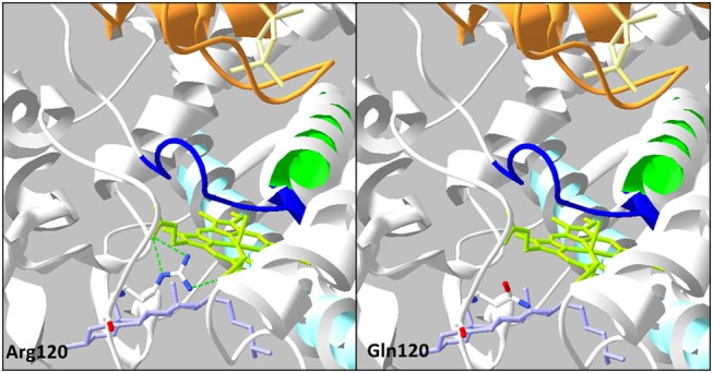
Analysis of the mutation p.Arg120Gln on three-dimensional model of CYP11A1 (PDB: 3N9Y). This amino acid replacement leads to disruption of three H-bonds (green dotted line) with the heme in green anis. The cholesterol is in violet, the I helix in light blue, L helix in green, cystein pocket in deep blue, and ferrodoxin in orange.