
Figure 5.
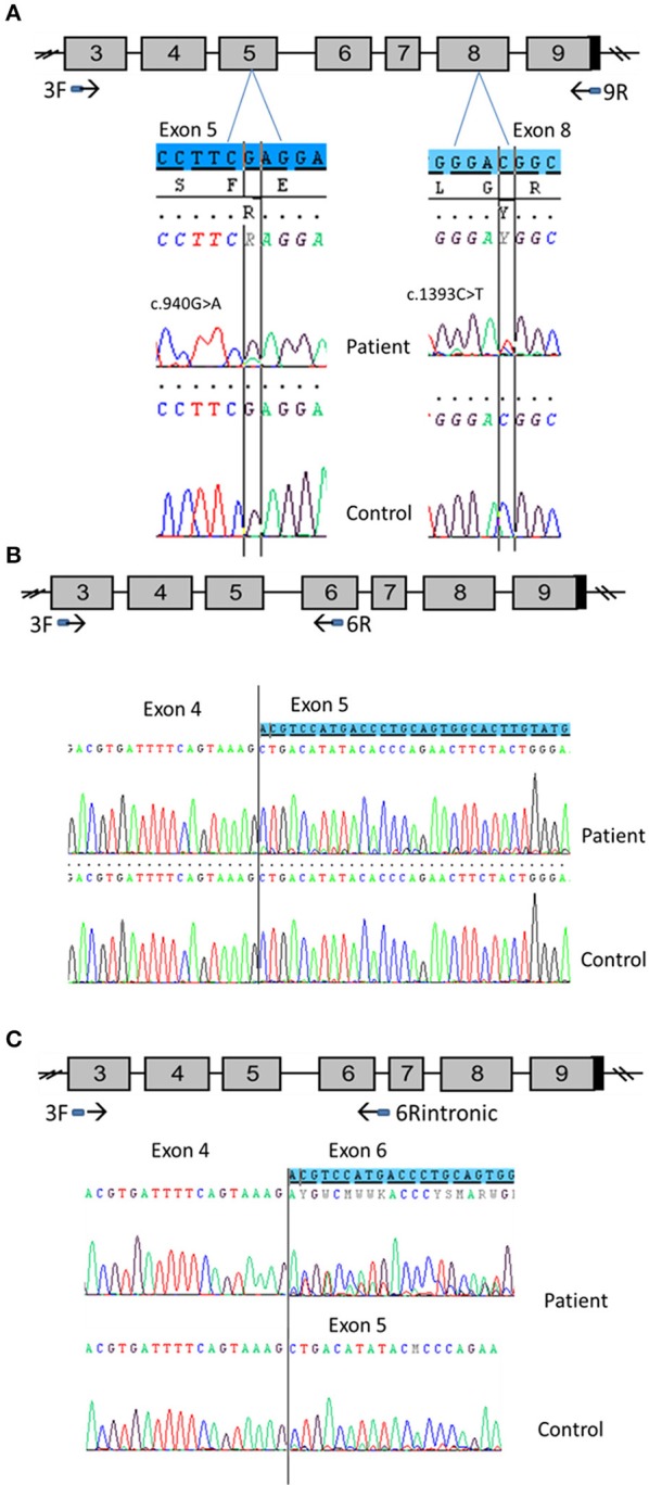
CYP11A1 mRNA analysis from testicular tissue of patient 1 compound heterozygous for the p.Glu314Lys and p.Arg465Trp mutations. (A) Amplification of CYP11A1 exons 3–9 (the reference sequences of exon 5 and 8 are highlighted in blue). Sequences of patient cDNA found the c.940G>A mutation in exon 5 and the c.1393C>T mutation in exon 8 at heterozygous state. WT nucleotide is more important than mutated nucleotide in exon 5 and in contrast, there was more mutated than WT nucleotide in exon 8. (B) Amplification of CYP11A1 exons 3–6. Sequence of patient exon 5 found a low-level second sequence, which matches sequence of exon 6 (the reference sequence of exon 6 is highlighted in blue). This is not the case for the control sample (underneath). (C) Amplification of exon 3 to intron 6. Sequence of patient cDNA is above and shows the skipping of exon 5 at heterozygous state (the reference sequence of exon 6 is highlighted in blue). Sequence of WT, underneath, shows this skipping at a very low level.