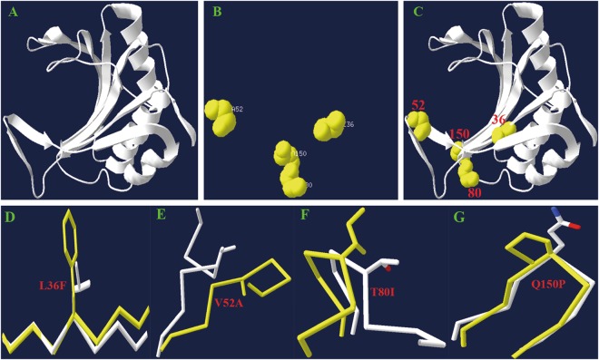
Figure 4.
Analysis of BraA.eIF(iso)4E.c-1 protein domain tertiary structure. (A) The three-dimensional structural model was built based on the wheat eIF(iso)4E protein. (B) Four amino acid variations were detected between the BraA.eIF(iso)4E.c-1 and BraA.eIF(iso)4E.c-2 proteins. (C) The location of the three different amino acid variations is depicted in the structural model of the entire protein. (D) The 36th amino acid changed from phenylalanine to leucine. (E) The 52th amino acids changed from valine to alanine. (F) The 80th changed from isoleucine to threonine. (G) The 150th changed from proline to glutamine.

