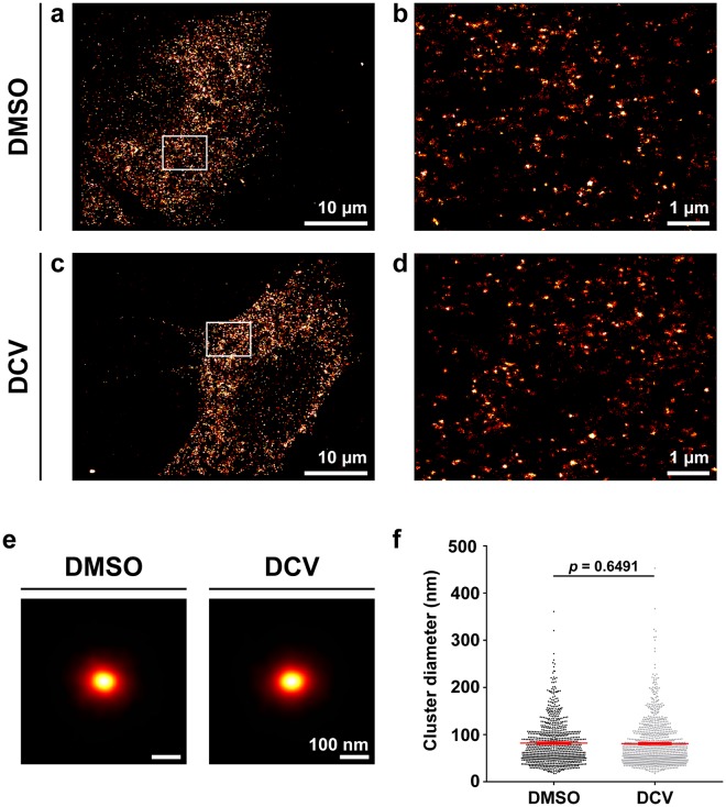
Figure 7.
DCV treatment of JFH-1 infected cells does not change NS3 cluster sizes. 3D-dSTORM images of Huh7 cells 24 hours post-infection with JFH-1, treated for 8 h with either DMSO or DCV. (a,c) Projections of 2 µm-thick volumes, 100 nm histogram bins. (b,d) Regions of interest within (a,c). Images are sum projections over depth (z) smoothened with a Gaussian filter σ = 20 nm. (e) NS3 cluster diameters. Mean and standard error of the mean are shown. Statistical significance (not significant) determined by two-tailed Student’s t-test with Welch’s correction.

