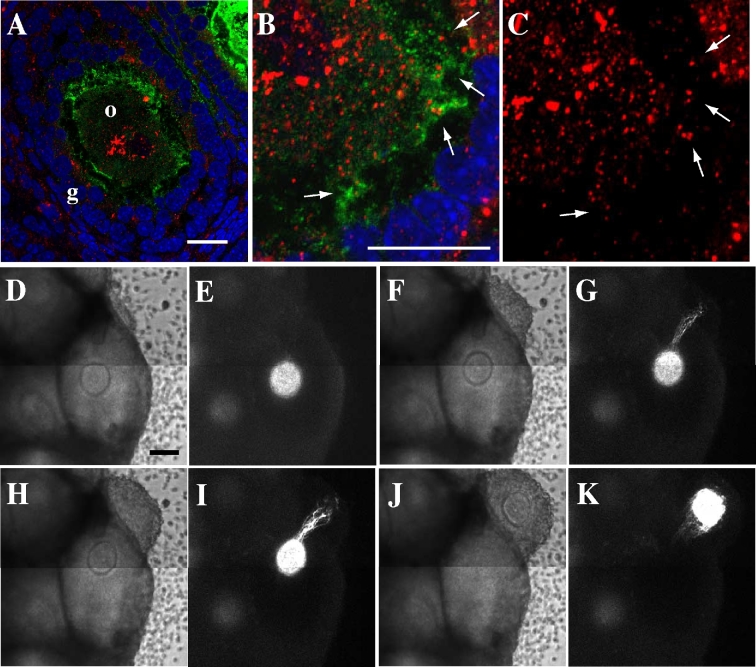
Figure 6.
The role of CCOC in follicle development. (A–C) Secondary follicle images stained with anti-GFP antibody (green), anti-GDF-9 antibody (red), and DAPI (blue). The arrows in (B) and (C) denote localization of GDF-9 in AcGFP1-positive CCOCs. (D–K) Time-lapse images of a cultured ovary in aOog1pro3.9 mouse. (D, F, H, J) Bright-field images. (E, G, I, K) AcGFP1 images. (D, E) 43 h culture. (F, G) 51 h culture. (H, I) 55 h culture. (J, K) 59 h culture. Notes: o, oocyte. g, granulosa cell. Scale bar, 100 μm.

