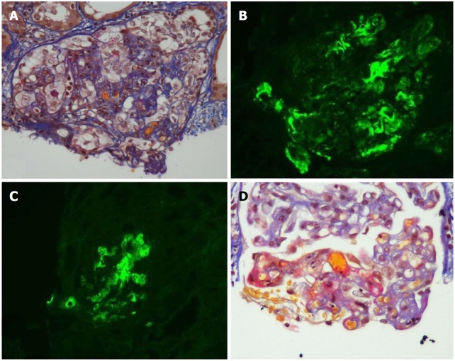
Figure 1.
Acute and chronic thrombotic microangiopathy and calcineurin inhibitors-associated arteriolopathy with severe acute ischemic tubular lesions. A: Advanced interstitial inflammatory fibrosis (Masson trichrome stain); B: Immunofluorescence, diffuse and segmental C3; C: C1q deposits within glomerular capillary walls; D: Diffuse acute and chronic arteriolar and glomerular thrombotic microangiopathy lesions on light microscopy (LM). (Adapted from: Yassine et al[45]).

