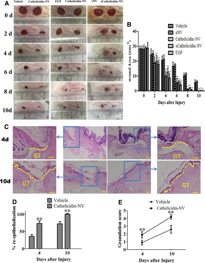
Figure 4. Topical application of cathelicidin-NV accelerated the healing of full-thickness wounds in mice.
(A) Images of a representative mouse from each group taken on post-injury days 0, 2, 4, 6, 8 and 10 are shown. Two full-thickness skin wounds of 6-mm diameter were created on the back of each mouse wound, and closure was monitored by the painted application of vehicle, cathelicidin-NV (20 μl, 20 µg/ml), EGF (20 μl, 100 µg/ml), sNV or sCathelicidin-NV (synthetic cathelicidin-NV). (B) Wound area at different days post injury. Values represent mean ± SEM (n = 10 per group). **P < 0.01 compared with control. (C) Images of skin tissue sections stained with H&E at days 4 and 10 post wounding. (The yellow dotted line: the neo-epithelium, GT: indicates granulation tissue, Scale bar = 100 µm). (D) Percent re-epithelialization at days 4 and 10 post wounding. (E) Histological scores of granulation thickness at days 4 and 10 post wounding. Values represent mean ± SEM (n = 6 per group). **P < 0.01 compared with control.

