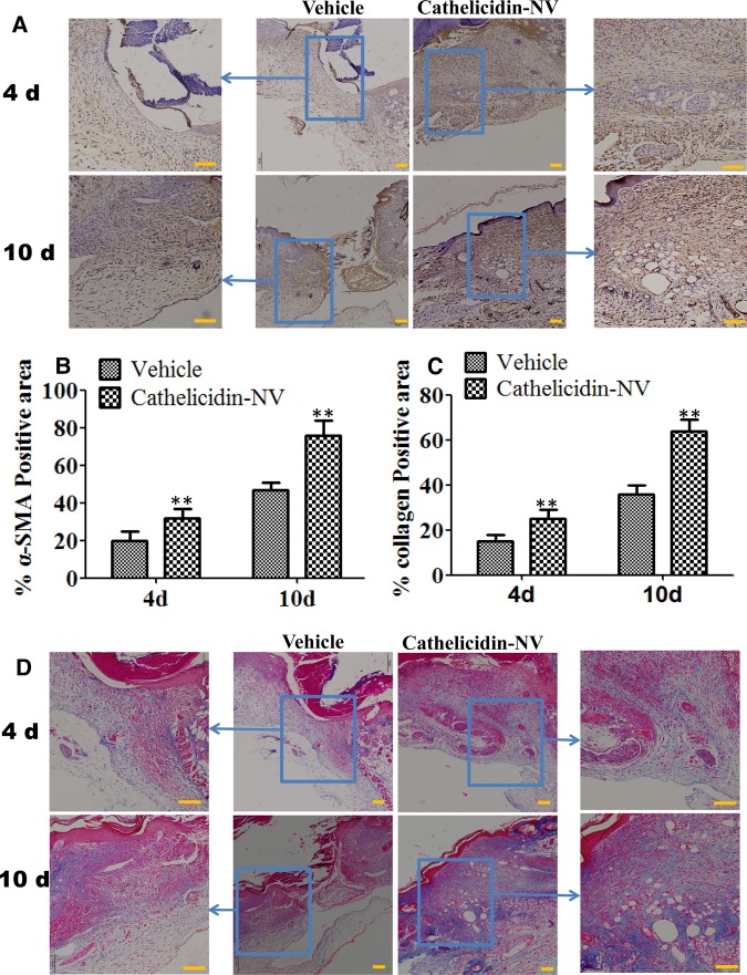
Figure 5. Cathelicidin-NV induced fibroblast-to-myofibroblast transition and significantly increased the production of collagen in the skin wound.
(A) The expression of α-SMA in wound skin treated with cathelicidin-NV or vehicle at days 4 and 10 after wounding. Myofibroblast are stained brown. (B) Quantification of α-SMA positive area. (C) Quantification of collagen positive area. (D) Histological assessment of collagen in the healing wounds. At days 4 and 10 post wounding, wounds with Vehicle and Cathelicidin-NV were stained for collagen with Masson Trichrome. Collagen bundles are stained blue. Scale bar = 200 µm. The IPLab imaging software were used to measure the changes of the wound. All values represent mean ± SEM (n = 6 per group). **P < 0.01 compared with control.

