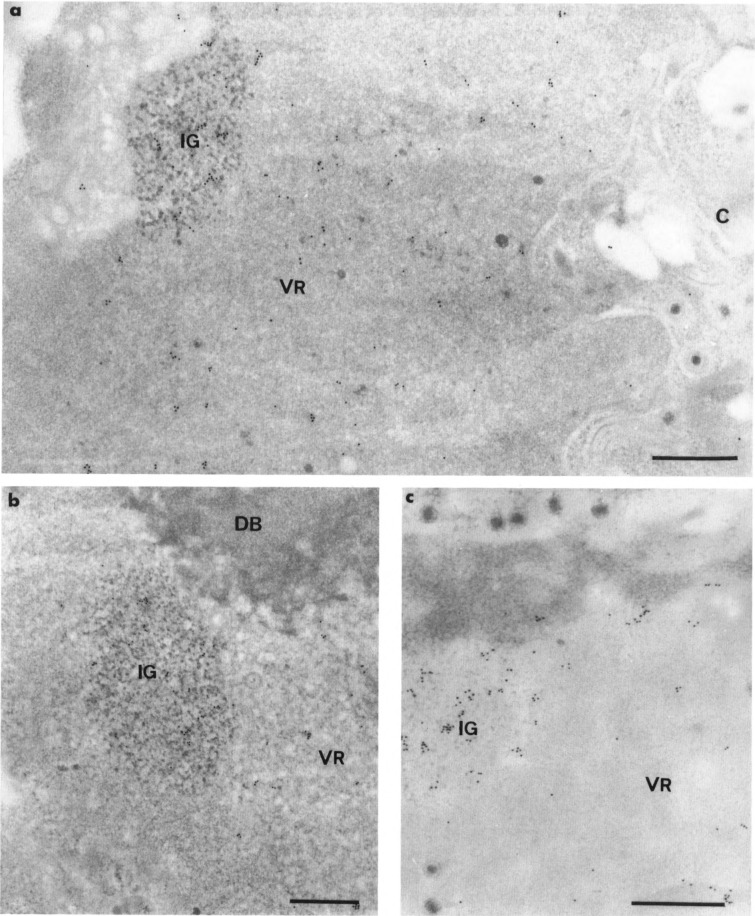
FIG. 7.
Localization of U1 RNA in HSV-1-infected HeLa cells by in situ hybridization of an U1 DNA probe. Formaldehyde fixation and Lowicryl K4M embedding. Uranyl acetate staining. (a,b) Nondigested sections. Gold particles are numerous in the nucleus over the clusters of interchromatin granules (IG) and the virus-induced regions (VR). In (a), a few gold particles also are present in the cytoplasm (C) over its ribosome-rich areas whereas the viruses are devoid of labeling. In (b), the virus-induced intranuclear dense body (DB) is not labeled, (c) Protease pretreatment of section prior to hybridization. Elimination of the proteins of the section does not modify the intensity and the distribution of labeling. Gold particles are still associated with the intranuclear virus-induced region (VR) and the cluster of interchromatin granules (IG). Bars represent 0.5 μm.

