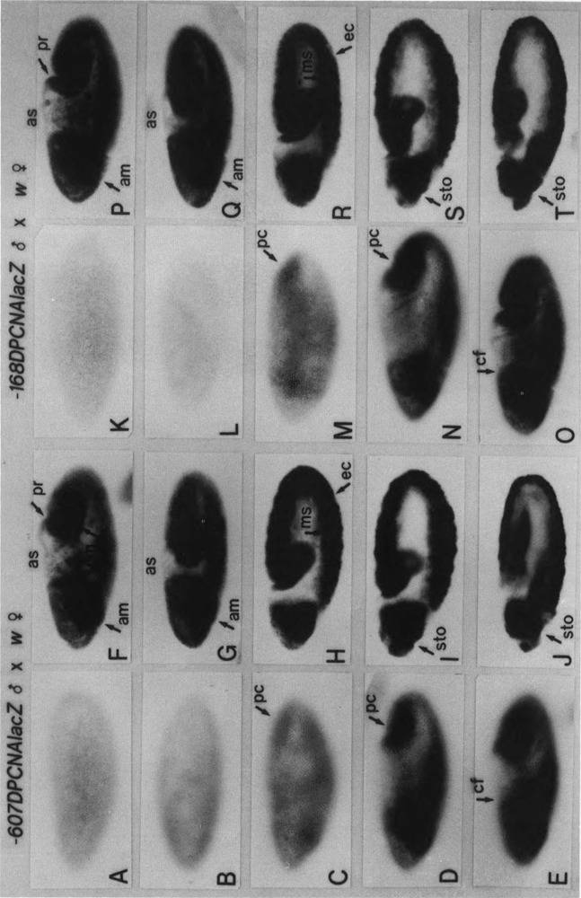
FIG. 3.

Zygotic expression of PCNA lacZ in embryos. Embryos from p5′-607DPCNAlacZW8HS males × w females (A–J) or p5′-168DPCNAlacZW8HS males × w females, as indicated at the top, were immunostained with an anti-lacZ antibody. Surface views of (A,K) syncytial blastoderm embryos (stage 4, 1320–1410 h), (B,L) cellular blastoderm embryos (stage 5, 1410–1450 h), (C,M) early gastrulating embryos (stage 6, 1450–1500 h), (D,N) embryos at the beginning of germ band elongation (early stage 7, 1500–1510 h), and (E,O) embryos at the early phase of germ band elongation (late stage 7) are shown; pole cells (pc) and cephalic furrow (cf). (F,P) Internal focal views of embryos shown in (E,O), anterior midgut primordium (am), proctodeum primordium (pr), posterior midgut primordium (pm), and aminoserosa (as). Internal focal views of (G,Q) stage 8 embryos (1510–1540 h), (H,R) stage 9 embryos (1540–1620 h), (I,S) stage 10 embryos (1620–1720 h), and (J,T) stage 11 embryos (1720–1920 h) are shown; mesoderm (ms), ectoderm (ec), stomodeum (sto).
