
FIG. 4.
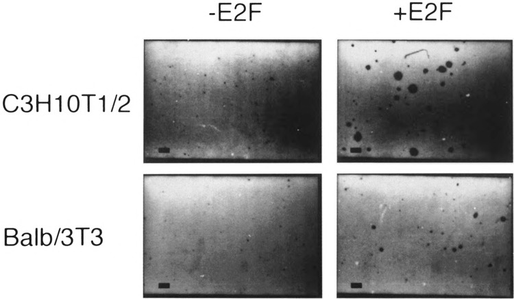
Transformation of C3H10T1/2 and Balb/3T3 clone A31 cells. Cells were plated in soft agar medium, stained, and photographed. Identity of cells is indicated to the left of the pictures and whether they overexpress E2F-1 is shown on the top. Magnification is ×40. The bar at the lower left corner of each micrograph is 200 μm.