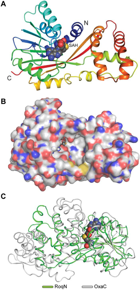
Fig. 4. RoqN crystal structure.
(A) Cartoon representation of RoqN shown in rainbow coloring. Bound SAH is shown as spheres. (B) Surface representation colored by atom of RoqN monomer. The active site is large and open to solvent. (C) Alignment of RoqN (green) and OxaC (gray) methyltransferases. Differences between the acceptor binding domains are apparent, while the SAM binding domains are conserved.

