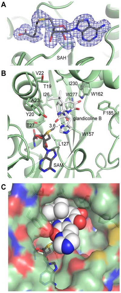
Fig. 5. RoqN substrates and modeling.
(A) Bound SAH in the RoqN active site. The electron density (3.0σ) was calculated from coefficients of the form Fo - Fc where SAH was omitted from the phase calculation and refinement. (B) Glandicoline B (white) was modeled into the RoqN active site (green) using Autodock Vina. SAM was modeled manually using the electron density for SAH followed by real space refinement. The lowest energy dock (−9.4 kcal/mol, shown) gave a close distance (3.6Å) between the donor methyl and the acceptor hydroxylamine in the expected near linear arrangement. The conserved Tyr20 is not positioned to activate the acceptor hydroxylamine. (C) Space-filling representation shows the open nature of the RoqN active site.

