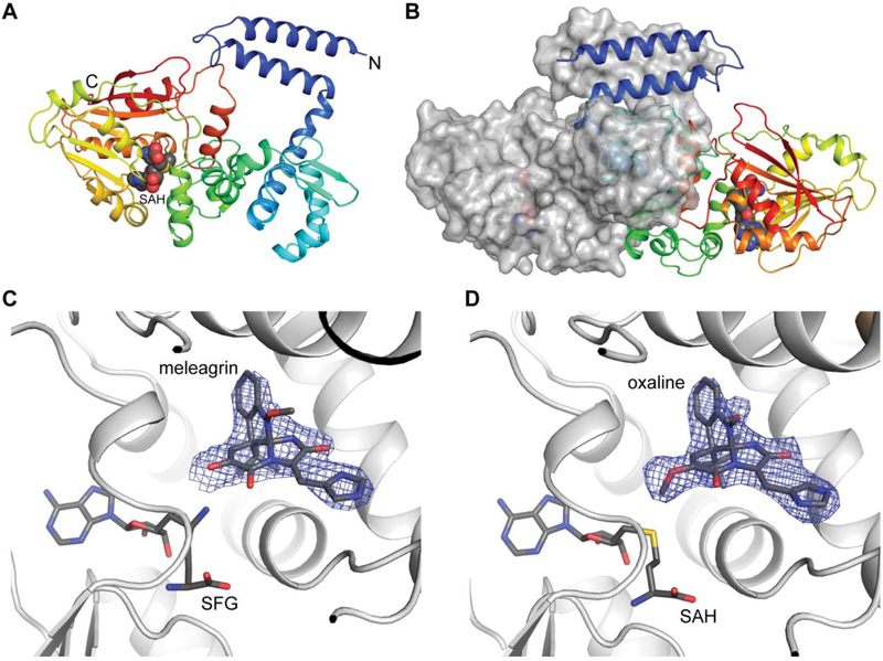
Fig. 6. OxaC complex structures.
(A) Cartoon representation of OxaC monomer shown in rainbow. SAH is shown as spheres. (B) OxaC dimer depicted as a hybrid of cartoon and surface representation. The two N-terminal helices (blue) form a four-helix bundle in the dimer. (C) Pseudo-Michaelis complex with OxaC·sinefungin (SFG)·meleagrin. (D) Product complex with OxaC·S-adenosyl-L-homocysteine (SAH)·oxaline. The electron density (Fo - Fc, 2.5σ contours) was calculated from a structure where the acceptor was omitted from the phase calculation and refinement.

