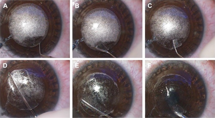
Figure 3.
Lenticule dissection and extraction.
Notes: (A) Cap side cut opened with hooked instrument. (B) Anterior lamellar plane delineated on the left-hand side. (C) Posterior lamellar plane delineated on the right-hand side. Meniscus sign confirms the identification of lenticule edge. (D) Anterior plane was dissected first. (E) Posterior plane dissection. (F) Lenticule extracted via microforceps.

