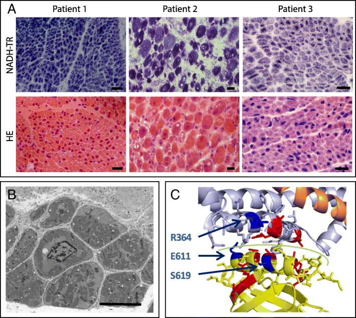
Fig. 1.
a. Hematoxilin-eosin (HE) and nicotinamide adenosine dinucleotide-tetrazolium reductase (NADH-TR) staining of muscles from the patients, showing fibers with centralized nuclei (HE) and abnormal central accumulation oxidative staining and a paler peripheral halo. Scale bars 20 μm. b. Electron microscopy of patient 1 muscle showing partial sarcomeres disorganisation and central nuclei. Scale bar 10 μm. c. Localization of presently reported mutations (dark blue) compared to known DNM2-CNM mutations (red) on the 3D model of nucleotide-free human DNM1 (PDB 3SNH). They all clusterize at the PH (yellow) – Middle/stalk (light blue) interface (green line)

