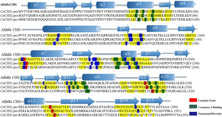
Fig. 4.
Structure and activity sites of the three CHS genes obtained from citrus
This figure was drawn according to Austin and Noel [37]. Key sections are highlighted in yellow. The CHS catalytic triad, residues bound to CoA, and other residues important for functional diversity are highlighted in red, green, and blue, respectively. For clarity, only identical residues in the equivalent positions of the aligned sequences are highlighted

