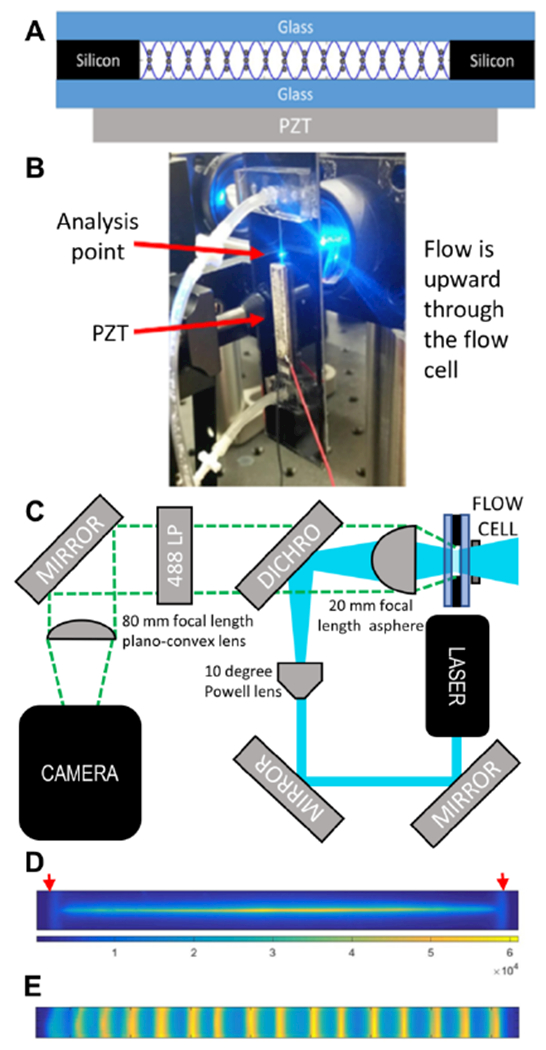
Figure 1. System description.

(A) Cross-sectional schematic of the acoustic focusing flow cell. The standing acoustic pressure wave (sine wave) focuses particles to the 16 pressure nodes across the width of the flow cell. (B) Image of the mounted flow cell. (C) Schematic of optics. (D) Laser excitation profile. The red arrows show the positions of the walls, that were 2.3 mm apart. (E) Stacked image of acoustically focused fluorescent particles.
