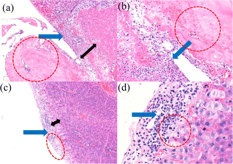
Fig. 6.

Histopathological examination with HE staining observed under a, c magnification × 100 and b, d magnification × 200. Histopathological examination shows an inflammatory response and liver injury among hepatocytes in two groups at the seventh day. The two groups are the a, b airflow-assisted group and c, d electric field-modified group (blue arrow: inflammatory cells; red circle: medical glue; black arrow: thickness of hyperemia zone)
