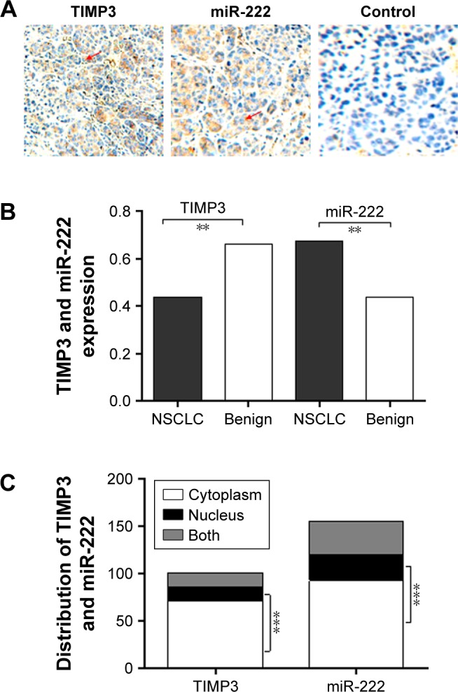
Figure 1.

Expression and distribution of TIMP3 and miR-222 in lung cancer.
Notes: The results of the IHC test were as follows: (A) PBS was used as a negative control, and the microscope was observed ×200 times. The protein expression score was evaluated according to the method. (B) The expression of miR-222 in lung cancer tissues was significantly higher, while that of TIMP3 was lower than that in normal lung tissues. (C) TIMP3 and miR-222 were mainly distributed in the cytoplasm (arrow). Scale bar = 50 µm; magnification ×20. **P<0.001; ***P<0.0001.
Abbreviations: IHC, Immunohistochemistry; miR-222, microRNA-222; NSCLC, non-small cell lung cancer; TIMP3, matrix metalloproteinase inhibitor 3.
