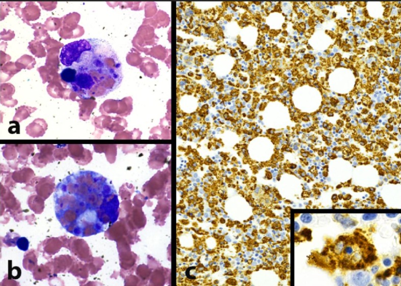
Figure 1.

Morphologic evidence of hemophagocytosis: bone marrow pathology pictures. (a) Macrophage with intracytoplasmic nuclear debris, red blood cells, and platelets (hemophagocytosis) (BM aspirate smear, Wright Giemsa stain, 1,000 × magnification). (b) Macrophage with numerous intracytoplasmic red blood cells (erythrophagocytosis) (BM aspirate smear, Wright Giemsa stain, 1,000 × magnification). (c) Extensive macrophage infiltration of bone marrow, highlighting scattered hemophagocytic cells (inset, 1,000 × magnification) (BM trephine core biopsy, CD68 immunohistochemical stain).
