Figure 1.

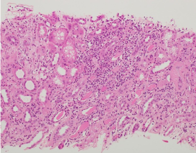
Light microscopy of kidney biopsy specimen (stained with H&E) showed tubulointerstitial inflammation: interstitial inflammatory infiltrates with prominent granular sites including eosinophils and neutrophils.

Light microscopy of kidney biopsy specimen (stained with H&E) showed tubulointerstitial inflammation: interstitial inflammatory infiltrates with prominent granular sites including eosinophils and neutrophils.