Figure 2.
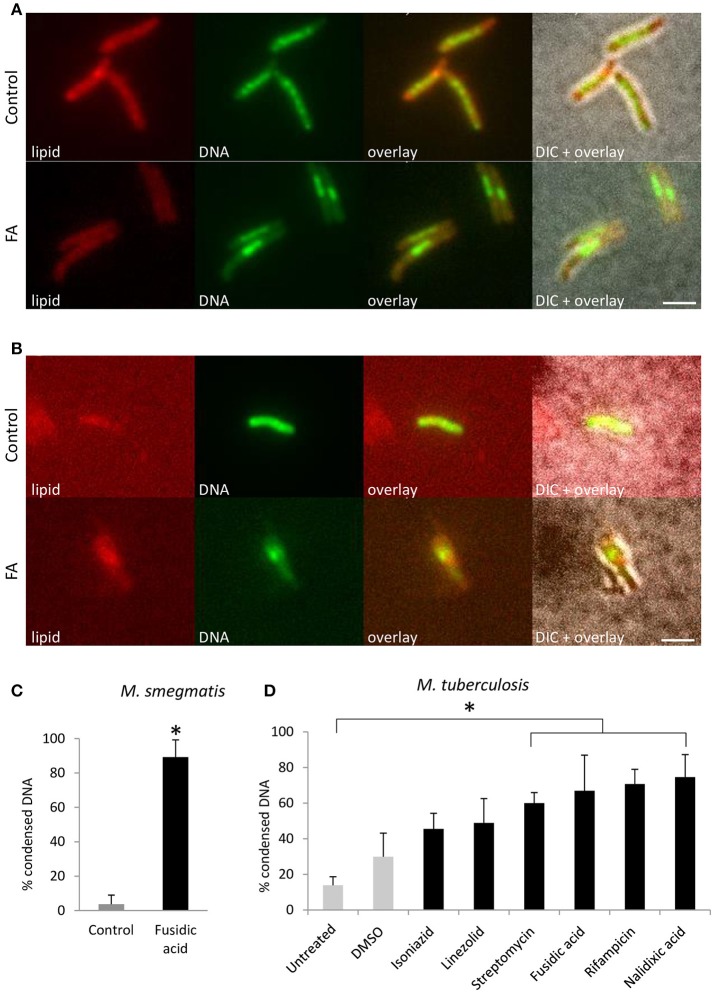
DNA condensation is a generic response to antibiotic-induced stress in Mycobacterium. Fluorescent microscopy images of lipid and DNA patterns in M. smegmatis mc2155 (A) and M. tuberculosis mc26030 (B) in control and FA treatment conditions. Lipids stained with BODIPY (red), DNA stained with Hoechst 33342 (green), an overlay of the two fluorescent signals and the overlay with the bright-field image (DIC). (C) Average bacteria with condensed DNA distribution patterns in M. smegmatis, in control (gray bar) and after 1 h FA-treatment (black bar, bars represents mean ± standard error, n = 3, *P < 0.05). (D) M. tuberculosis mc26030 was exposed to various antibiotics to target different cellular processes. The incidence of DNA condensation was quantified for untreated, DMSO treated as a control (gray bars) and antibiotic treated (black bars) M. tuberculosis mc26030 cultures. Per condition, the percentage of bacilli with condensed DNA is displayed, which was based on three measurements of n ≥ 20 bacteria per condition. Treatment with streptomycin, fusidic acid, rifampicin and nalidixic acid increased the percentage bacteria with condensed DNA significantly (bar represents mean ± standard error, *P < 0.05). Scale bar represents 2 μm.

