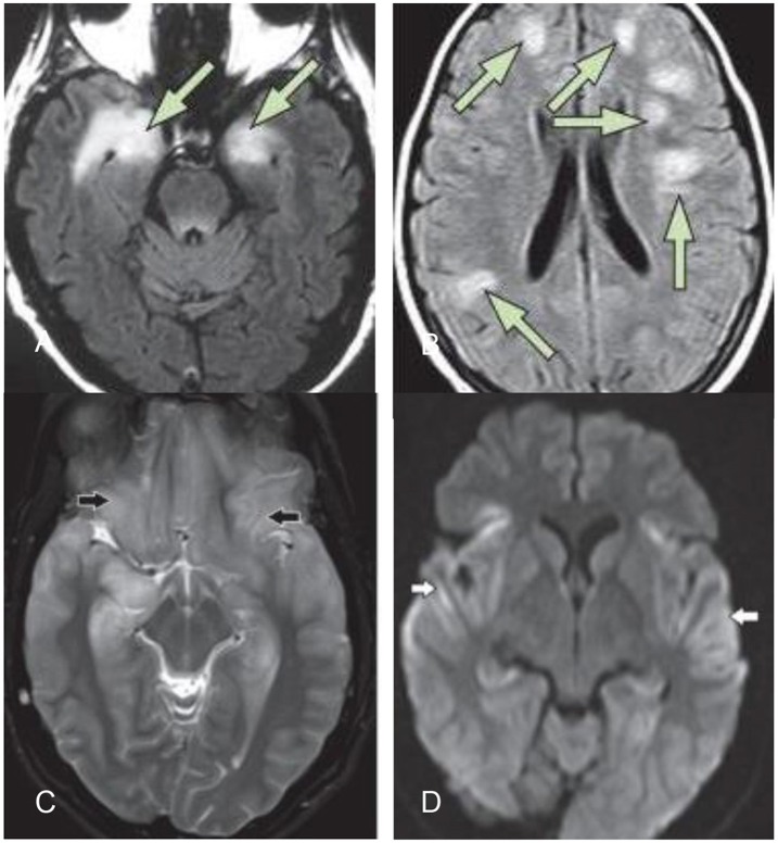
Figure 1.
(A) Typical MRI of limbic encephalitis with bilateral hyperintensities in the medial temporal lobe on T2-weighted fluid–attenuated inversion recovery imaging (B) typical MRI of ADEM (14). (C,D) Herpes simplex virus encephalitis: bilateral symmetric cortical swelling and hyperintensity on T2 weighted image involving the anteromedial temporal lobes, insular cortex, orbital gyri (black arrows) with restricted diffusion in the involved areas (white arrows) (25).

