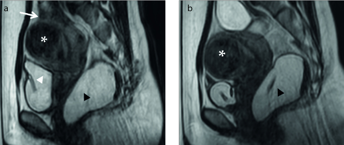
Figure 13. a, b.
Sagittal T2-weighted images show the uterine fibroid (asterisk), bowel, bladder and rectum. In panel (a), the bladder filled with 300 mL normal saline (white arrowhead) and rectum filled with 200 mL ultrasound gel (black arrowhead) pushes the uterus upward and forward. The bowel loops are still above the tumor (white arrow). Meanwhile in panel (b), the bladder is emptied with the pressure of the filled rectum. The uterus moves forward and downward and the bowel loops are pushed out of treatment window.

