
Figure 1.
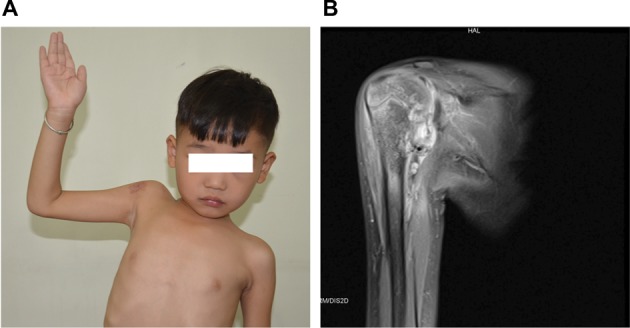
Deep KHE with bone–joint erosion.
Notes: (A) A 6-year-old boy with decreased right shoulder ROM for 3 years. (B) Coronal T2-weighted MRI shows a deep lesion with marked abnormal enhancement at the proximal humerus, glenoid cavity, scapula, and the surrounding soft tissue.
Abbreviations: KHE, kaposiform hemangioendothelioma; MRI, magnetic resonance imaging; ROM, range of motion.