
Figure 2.
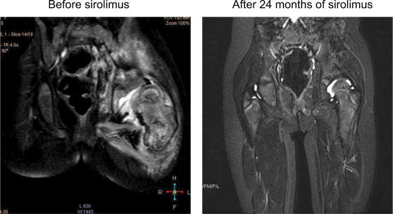
MRI changes in KHE with decreased hip ROM before and at 24 months after treatment.
Notes: A 2.2-year-old girl demonstrated progressively decreased hip ROM for 12 months (left). Coronal T2-weighted MRI revealed a deep lesion infiltrating the proximal femur and ilium. MRI at 24.0 months after sirolimus treatment revealed nearly complete involution of the lesion (right).
Abbreviations: KHE, kaposiform hemangioendothelioma; MRI, magnetic resonance imaging; ROM, range of motion.