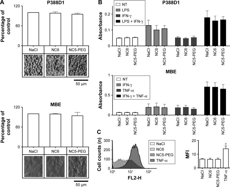
Figure 3.
Analysis of inflammatory response to nanocapsules.
Notes: (A) Viability of mouse macrophage cell line P388D1 and endothelial MBE cells. The cells were incubated for 48 hours in growth medium with 1.5 mM NaCl or nanocapsules (~2×10¹¹/mL). (B) Analysis of NO production by iNOS. Nitrite levels were measured by Griess reaction after 24 hours incubation of the cells with 1) nanocapsules or NaCl (NT), 2) LPS or cytokines and 3) nanocapsules and LPS or cytokines. (C) Flow cytometry analysis of VCAM-1 expression on MBE cells. MBE cells were incubated for 24 hours with nanocapsules or with TNF and then stained with anti-VCAM-1 antibody. *P<0.05
Abbreviations: LPS, lipopolysaccharide; MFI, mean fluorescence intensity; NC5-PEG, five-layer PEGylated nanocapsules; NC6, six-layer nanocapsules; NO, nitric oxide; PEG, polyethylene glycol; TNF, tumor necrosis factor; VCAM, vascular cell adhesion molecule.

