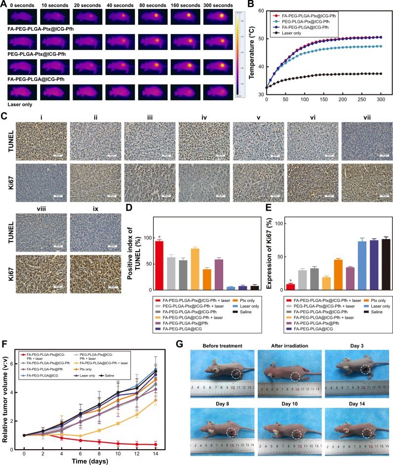
Figure 5.
In vivo anticancer therapy.
Notes: (A) Monitoring images of MDA-MB231 tumor-bearing mice in laser-irradiation groups during PTT by thermal imaging. (B) Temperature of tumor region in laser-irradiation groups during PTT. (C) TUNEL and Ki67 expression in tumor tissue by immunohistochemical staining. Nuclei, stained in brown, are from TUNEL-positive or Ki67-positive cells, with blue indicating negative (FA-PEG-PLGA-Ptx@ICG-Pfh NPs with laser irradiation [i], PEG-PLGA-Ptx@ICG-Pfh NPs with laser irradiation [ii], FA-PEG-PLGA-Ptx@ICG-Pfh NPs without laser irradiation [iii], FA-PEG-PLGA@ICG-Pfh NPs with laser irradiation [iv], FA-PEG-PLGA-Ptx@Pfh without laser irradiation [v], Ptx only [vi], FA-PEG-PLGA@ICG without laser irradiation [vii], laser only [viii], saline [ix]). (D) Positive index of TUNEL assay for FA-PEG-PLGA-Ptx@ICG-Pfh NPs with laser irradiation was higher than that in the other eight groups (P<0.05). (E) Ki67 expression for FA-PEG-PLGA-Ptx@ICG-Pfh NPs with laser irradiation was lower than that of the other eight groups (P<0.05). (F) Relative tumor-volume curves of different groups of MDA-MB231 tumor-bearing mice. (G) Typical MDA-MB231 tumor-bearing mice having received FA-PEG-PLGA-Ptx@ICG-Pfh with laser irradiation on different days. (H) Body weight of mice was measured during the 14 observation days in different groups. (I) H&E-staining images of major organs collected from the FA-PEG-PLGA-Ptx@ICG-Pfh NPs with laser irradiation and saline only (control) groups. *P<0.05 significant difference FA-PEG-PLGA-Ptx@ICG-Pfh compared with other groups.
Abbreviations: PTT, photothermal therapy; FA, folic acid; PEG, polyethylene glycol; PLGA, poly(lactide-co-glycolic acid); Ptx, paclitaxel; ICG, indocyanine green; Pfh, perfluorohexane; NPs, nanoparticles.


