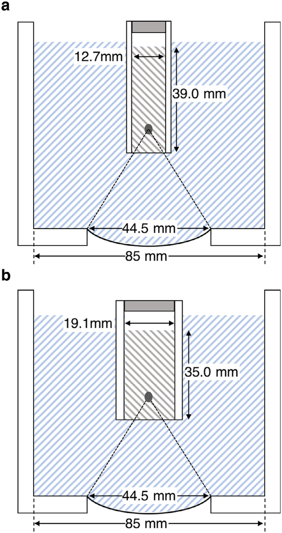
Fig. 1.
The transducer and water bath assembly for (a) 5-mL or (b) 10-mL samples. Although not strictly to scale, the location and size of the cavitation zone is approximately correct. A small air gap was left between the fixtures and the sample fluid to avoid displacing the bacterial suspensions when assembling the filled vessels.

