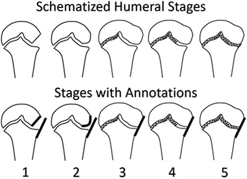
FIGURE 1.

Periphyseal changes around the proximal humerus: stage 1 demonstrates an incompletely ossified lateral epiphysis such that the lateral margin is oblique (short bold line). Stage 2 demonstrates increased ossification of the lateral epiphysis with a curvilinear lateral margin (bold curve). Note that in stages 1 and 2, the bold line parallel to the lateral metaphysis does not touch the epiphysis. Stages 3 through 5 all demonstrate colinearity between the lateral margin of the epiphysis and the metaphysis such that a single bold line touches both edges. In stage 3, the lateral half of the physis is open without obvious fusion. In stage 4, the lateral half of the physis thins and begins partial fusion. Finally, by stage 5 the lateral half of the physis demonstrates essentially complete fusion. Fusion is indicated by the hashes.
