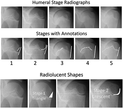
FIGURE 2.

Representative images of the humeral stages. Stage 1 demonstrates an incompletely ossified lateral epiphysis leaving a triangular area of radiolucency on the lateral aspect of the epiphysis. Stage 2 demonstrates increased ossification of the lateral epiphysis leaving a crescent shaped area of radiolucency on the lateral side of the epiphysis. These shapes are highlighted below the annotations with representative images shown both unmodified and with the shapes superimposed. Note that in stages 1 and 2, the black line parallel to the lateral metaphysis does not touch the epiphysis. Stages 3 through 5 all demonstrate colinearity between the lateral margin of the epiphysis and the metaphysis. In Stage 3, the lateral half of the physis is open without obvious fusion. In stage 4, the lateral half of the physis thins and begins partial fusion. Finally, by stage 5 the lateral half of the physis demonstrates essentially complete fusion. The same annotations used on the schematic are superimposed upon the radiographic examples for ease of comparison.
