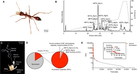
Fig. 1. The polypeptidic venom composition of M. gulosa.

(A) M. gulosa. (B) Total ion chromatogram of M. gulosa venom. Venom (2 μg) was separated by C18 ultrahigh-performance liquid chromatography (uHPLC), using a gradient of 1 to 40% solvent B [90% acetonitrile (ACN) and 0.1% FA] over 45 min and analyzed using MS/MS. Peaks corresponding to identified venom peptides are labeled. Peaks identified as corresponding to partial fragments of larger peptides are labeled with an asterisk. (C) Venom apparatus of M. gulosa. (D) Venom component-encoding transcripts (that is, those encoding peptides detected in the venom itself) comprised 15.9% of total expression. Of these, transcripts encoding aculeatoxin peptides, MIITX2-Mg1a, MIITX3-Mg1a, and venom proteins comprised 93.0, 3.4, 3.3, and 0.3%, respectively, of venom component expression. EGF, epidermal growth factor. (E) Venom component-encoding transcripts (highlighted in red) are found exclusively in the highly expressed portion of the venom apparatus transcriptome, where they constitute most of the most highly expressed transcripts. Inset: Zoom showing the top 50 transcripts (with a linear scale on the y axis). The most highly expressed nonvenom component-encoding transcripts are labeled [cytochrome c oxidase subunits 1 (COI), 2 (COII), and 3 (COIII)].
