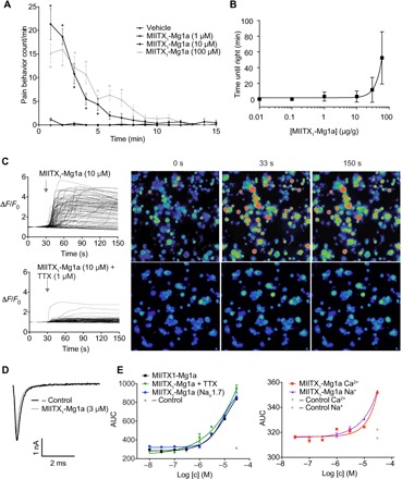
Fig. 4. Defensive and predatory function and mechanism of action of MIITX1-Mg1a.

(A) Intraplantar injection of MIITX1-Mg1a causes spontaneous dose-dependent nocifensive behavior in mice, lasting for up to 7 min (n = 6 per group). Data are expressed as means ± SEM. Statistical significance compared to vehicle control was determined using two-way analysis of variance (ANOVA) with Sidak’s multiple comparison test. *P < 0.05. (B) Intra-abdominal injection of MIITX1-Mg1a in crickets (A. domesticus) caused dose-dependent, reversible, and nonlethal incapacitation (inability to right after being placed on back). Effects were observed at doses ≥1 μg/g (0.3 nmol/g), and at the maximum dose tested (60 μg/g, 19 nmol/g), incapacitation lasted an average of 53 min. (C) MIITX1-Mg1a caused an increase in [Ca2+]i in DRG cells, which was mostly blocked in the presence of TTX. (D) Representative sodium currents obtained in HEK293 cells heterologously expressing hNaV1.7, in the presence and absence of 3 μM MIITX1-Mg1a. Currents were obtained by a 50-ms voltage pulse to −20 mV from a holding potential of −90 mV. Compared with buffer control, in hNaV1.7-expressing cells, MIITX1-Mg1a did not change peak current or activation/inactivation kinetics, and it did induce persistent currents. (E) In HEK293 cells [independent of hNaV1.7 expression or the presence of TTX (1 μM)], MIITX1-Mg1a alters membrane potential and causes Na+ and Ca2+ influx in a concentration-dependent manner. Concentration-response curves were evaluated by fitting the data with a four-parameter Hill equation with variable slope. FLIPR data are expressed as the means ± SEM and are representative of at least three independent experiments. AUC, area under the curve.
