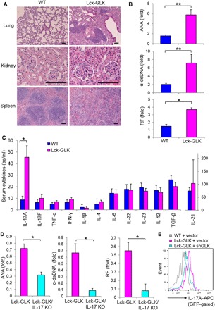
Fig. 1. Lck-GLK Tg mice display autoimmune phenotypes and selectively increased serum IL-17A levels.

(A) Hematoxylin and eosin (H&E)–stained sections of the indicated organs from 16-week-old mice. Scale bars, 100 μm. (B) Levels of serum autoantibodies from 20-week-old mice were determined by ELISAs. The levels are presented relative to the value from one of the wild-type (WT) mice. WT, n = 7; Lck-GLK, n = 8. (C) The serum levels of cytokines in 4-week-old mice were determined by ELISAs. WT, n = 20; Lck-GLK, n = 16. (D) The serum levels of autoantibodies in 20-week-old Lck-GLK and Lck-GLK/IL-17A KO mice were determined by ELISAs. The levels are presented relative to the value from one of the Lck-GLK mice. n = 6 per group. (E) IL-17A expression was attenuated by GLK shRNA. Murine primary splenic T cells were transfected with green fluorescent protein (GFP)–human GLK shRNA and a control GFP vector. The transfected T cells were stimulated with anti-mouse CD3 antibodies for 3 hours and then determined by flow cytometry at day 3 after transfection. Data show the events of IL-17A–producing T cells (GFP-gated). WT, wild-type littermate controls; Lck-GLK, T cell–specific GLK Tg mice; Lck-GLK/IL-17A KO, Lck-GLK;IL-17A–deficient mice; ANA, antinuclear antibody; α–double-stranded DNA (dsDNA), anti-dsDNA antibody; RF, rheumatoid factor; APC, allophycocyanin. Data shown are representative of three independent experiments. *P < 0.05, **P < 0.01 (two-tailed Student’s t test).
