
Fig. 4. Murine CTCs.
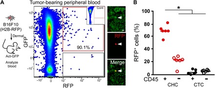
(A) B16F10 (H2B-RFP) cells (5 × 10⁴ cells) intradermally injected into a syngeneic GFP-expressing recipient mouse. Blood collected at time of tumor resection and analyzed by flow cytometry for GFP and RFP expression. RFP+GFP+ cells were detectible in presorted cell preparations by immunofluorescence. Scale bar, 50 μm. We analyzed GFP-expressing blood by flow cytometry as a negative control for (A) inset. (B) Percentages of fusion hybrids (RFP+/GFP+) and unfused CTCs (RFP+/GFP−) expressing the leukocyte antigen CD45 (*P < 0.000002).