
Fig. 5. Cell fusion in human tumors.
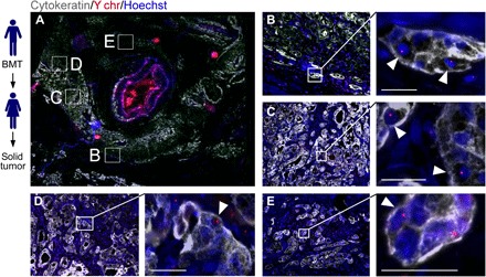
Solid tumors from women (n = 7) with previous sex-mismatched bone marrow transplantation (BMT) permits analysis of cell fusion. (A) PDAC tumor section with cytokeratin (gray), the Y chromosome (Y chr, red), and Hoechst (blue) detection revealed areas of cytokeratin-positive cells with Y chr–positive nuclei (white arrowheads). Boxed representative areas are enlarged in (B) to (E). Scale bars, 25 μm.