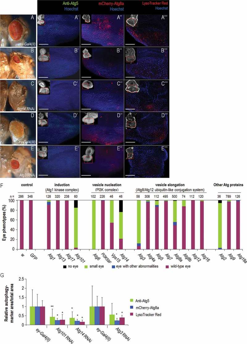
Figure 3.

Silencing of Atg genes in the eye disc can severely compromise eye morphology in the affected adults. (A) Stereomicrograph of an ey-Gal4(II)/+ adult head, which served as a control for silencing of Atg101 and Atg14, showing wild-type eye morphology. (A’) Atg5 antibody staining indicates early autophagic structures (green dots) in the eye disc of an ey-Gal4(II)/+control animal. (A’’) mCherry-Atg8a accumulation in the eye disc of an ey-Gal4(II)/+ control animal. Fluorescent foci (red) indicate autophagosomal and autolysosomal structures. (A’’’) LysoTracker Red staining marks acidic structures in the eye disc of an ey-Gal4(II)/+ control animal. (B) Small eye phenotype of an ey-Gal4(II)/Atg101 RNAi adult. (B’ to B’’’) Silencing of Atg101 in the eye disc leads to reduced levels of Atg5- (B’) mCherry-Atg8a- (B’’) and LysoTracker Red- (B’’’) positive structures. (C) The eyeless phenotype of an ey-Gal4(II)/Atg14 RNAi adult. (C’ to C’’’) Depletion of Atg14 in the eye disc leads to reduced levels of Atg5- (C’) mCherry-Atg8a- (C’’) and LysoTracker Red- (C’’’) positive structures. (D) Stereomicrograph of an ey-Gal4(III)/+ adult head, which served as a control for silencing Atg3, showing wild-type eye. (D’) Atg5 accumulation, (D’’) mCherry-Atg8a expression and (D’’’) LysoTracker Red staining in the ey-Gal4(III)/+ genetic background. (E) Stereomicrograph of an ey-Gal4(III)/Atg3 RNAi adult head showing a small eye phenotype., (E’) Silencing of Atg3 in the eye disc leads to a reduced amount of Atg5-, (E’’) mCherry-Atg8a-, and (E’’’) LysoTracker Red-positive foci. In images A’ to A’’’, B’ to B’’’, C’ to C’’’, D’ to D’’’ and E’ to E’’’, the antenna part is up; bars: 50 µm. At the upper left corner of each image, the red rectangle indicates the area enlarged. Eye disc samples were dissected from L3W larvae. (F) Silencing of Atg genes in the eye primordium can severely compromise the development of the organ. The penetrance of eye phenotypes may depend on the efficiency of the RNAi constructs used (also see Figs. S9 and S10). In some cases, like Atg2, Atg3, Atg6, Atg14 and Atg101, the phenotype is manifested with a nearly full penetrance. (G) Effect of Atg3, Atg14 and Atg101 RNAi treatments on autophagic activity in the eye disc of L3W larvae. The ratio of anti-Atg5/mCherry-Atg8a/LysoTracker Red-positive structures and the area of entire eye disc in each image is shown as averages, the data represent relative values. Bars represent mean ±S.D., *: P < 0.05, **: P < 0.01, two-sample Student t test, t test for unequal variances or Mann-Whitney U test. Temperature: 29°C, with the exception of E’’: 18°C, and D to D’’’, E, E’, E’’’: 25°C. In fluorescence images, the background expression was highly reduced in order to strengthen the visibility of puncta.
