
Figure 6.
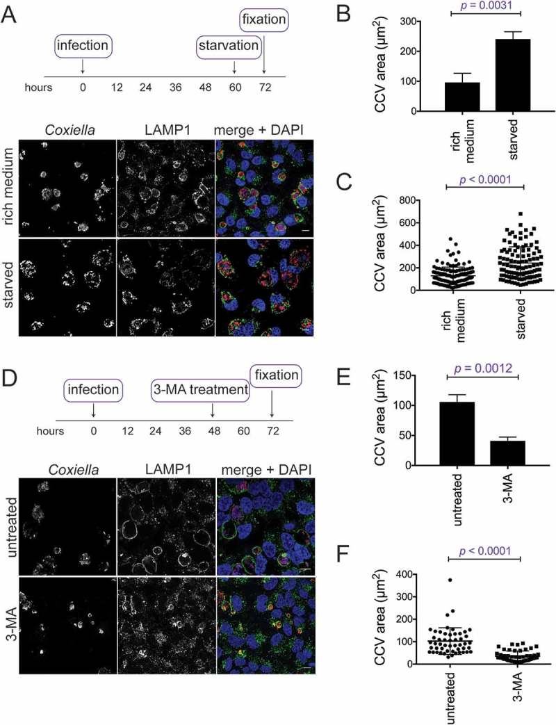
CCV size depends on the autophagic state of the host cell. (a) HeLa cells infected with WT Coxiella for 60 h were starved in HBSS for 12 h before being fixed and stained with anti-Coxiella (red), anti-LAMP1 (green), and DAPI (blue). Images are representative of CCV formation. Scale bar: 10 µm. CCV areas were (b) measured over 3 experiments, or (c) displayed as individual data points (n = 50) from 1 experiment. Error bars represent SD. (d) HeLa cells were infected with WT Coxiella for 48 h and treated with 10 mM of 3-MA for 24 h, or left untreated. Samples were then fixed and stained with anti-Coxiella (red), anti-LAMP1 (green), and DAPI (blue). Scale bar: 10 µm. CCV areas were measured (e) over 3 experiments, or (f) displayed as individual data points (n = 50) from 1 experiment. Error bars represent SD.