
Figure 7.
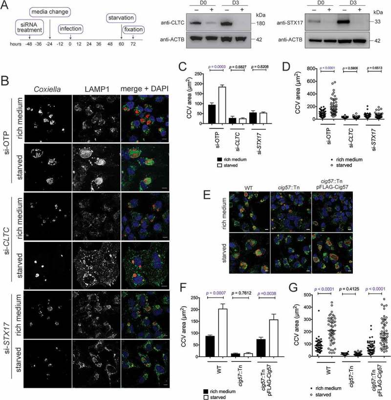
Autophagy-dependent CCV expansion requires CLTC. (a) Immunoblots showing CLTC or STX17 and ACTB during respective siRNA treatments (+) or non-targeting treatment (–), where samples were taken at the day of infection (D0, 2 days post siRNA treatment) and 3 days post infection (D3). (b) Confocal images of HeLa cells, treated with non-targeting (si-OTP) siRNA, siRNA targeting CLTC (si-CLTC), or STX17 (si-STX17) before being infected with Coxiella for 60 h and then incubated in either normal DMEM + 10% FBS (rich medium) or in HBSS starvation medium (starved) for 12 h before fixation. Samples were stained with anti-Coxiella (red), anti-LAMP1 (green), and DAPI (blue) to highlight CCV formation. Scale bar: 10 µm. (c) Quantification of CCV sizes during the above conditions. Data represents the average CCV area and SD over 3 independent experiments. (d) Individual data points from 1 experiment quantifying CCV size (n = 50) to show the spread of the data. (e) Representative confocal images of HeLa cells infected with WT, cig57::Tn, or cig57::Tn pFLAG-Cig57 Coxiella for 60 h before starvation treatment for 12 h. As above, anti-Coxiella (red), anti-LAMP1 (green), and DAPI (blue) highlight the formation of CCVs. Scale bar: 10 µm. (f) Quantification of CCV area over 3 independent experiments shown in E. Error bars represent SD. (g) Individual data points (n = 50) from 1 experiment showing the observed variation in CCV size. Error bars represent SD.