
Figure 7.
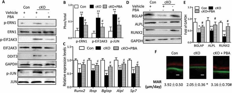
Administration of PBA partially reversed the suppression of bone formation by Atg7 deletion in vivo. PBA (5 mg/Kg) or vehicle was daily intraperitoneal injected to 3-wk-old cKO or control mice for 4 wks. By the end of 4-wk treatment period, total protein and RNA was extracted from calvaria (n = 3/group). (a and b) Expression of the ER stress markers (p-ERN1, p-JUN, DDIT3 and p-EIF2AK3). (c) Relative expression of Runx2, Ibsp, Bglap, Alpl, and Sp7 mRNA normalized to controls. (d and e) Expression of bone formation markers (BGLAP, ALPL and RUNX2). (f) Representative images of the double label staining in femur section with calcein and xylenol orange. Scale bars: 10 µm (mean ± SD, one-way ANOVA, *P < 0.05, **P < 0.01 cKO vs. Con, # P < 0.05 cKO + PBA vs. cKO).