
Figure 8.
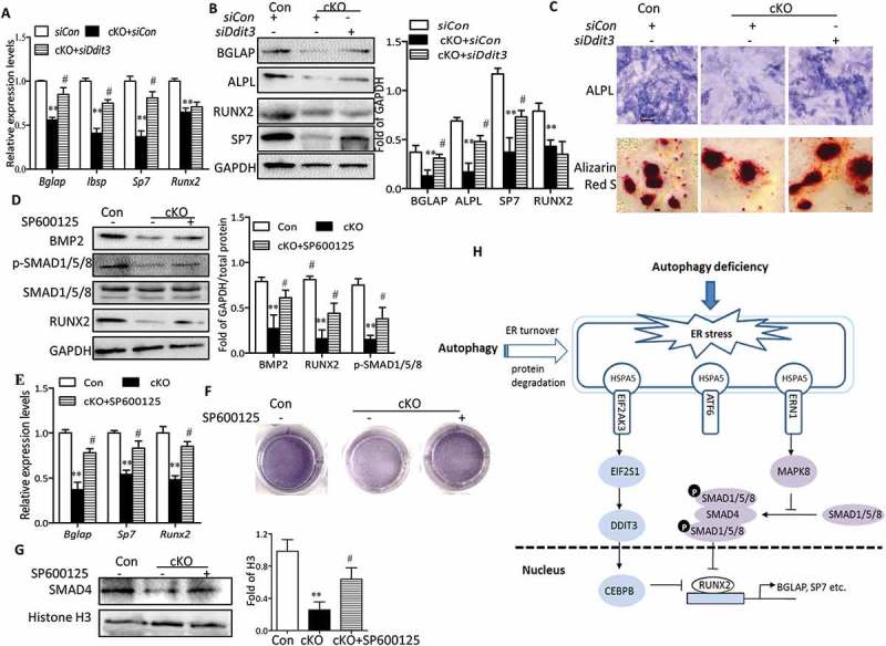
Autophagy deficiency impedes osteoblast differentiation partially via DDIT3- and MAPK8-SMAD1/5/8-dependent signaling. Relative expression of Bglap, Sp7, Ibsp and Runx2 mRNA (a), and BGLAP, ALPL, SP7 and RUNX2 proteins (b) were examined by RT-qPCR or western blot in primary osteoblasts transfected with or without siDdit3. Values expressed as mean ± SD of 3 independent experiments (one-way ANOVA, **P < 0.01 cKO + siCon vs. siCon, # P < 0.05 cKO + siDdit3 vs. cKO + siCon). (c) Representative images for ALPL and Alizarin red S staining in osteoblasts. Scale bars: 100 µm. Primary osteoblasts were pretreated with SP600125 (10 µM) for 2 h and then cultured in differentiation medium for 7 days. Representative western blot of BMP2 and RUNX2 and phosphorylation of SMAD1/5/8 (d), RT-qPCR for Bglap, Sp7 and Runx2 mRNA (e), representative images for ALPL staining (f), and representative western blot of nuclear SMAD4 expression (g) in osteoblasts, representative of 3 experiments (mean ± SD, one-way ANOVA, *P < 0.05 cKO vs. Con, # P < 0.05 cKO + SP600125 vs. cKO). (h) Model depicting the mechanism through which autophagy deficiency-mediated ER stress leads to inhibited osteoblast differentiation.