Figure 7.

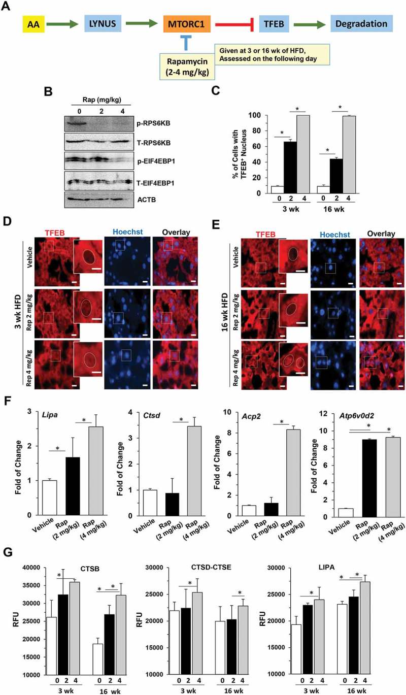
Suppression of MTORC1 disrupted the oscillation pattern of TFEB signaling and improved hepatic status in HFD-fed mice. (a) Experimental scheme. Mice fed with HFD for 3 or 16 weeks were given 2 mg/kg or 4 mg/kg rapamycin and sacrificed 16 h later. (b) Hepatic lysates were prepared from the 16-wk groups and analyzed by immunoblotting assay. The letter p indicates the phosphorylated form whereas T indicates all forms. (c-e) The percentage of cells with TFEB-positive nuclei (c) was quantified from livers stained with anti-TFEB antibody and Hoechst 33328 after 3-week (d) or 16-week (e) HFD feeding. Boxed areas were enlarged in the insets, which illustrate the representative hepatocytes. Dotted circles indicate the nucleus. (f) The mRNA levels of the selected TFEB targets were determined by qRT-PCR in mice fed with HFD for 16 weeks. (g) Hepatic lysosomal enzyme activities were determined. *, p < 0.05; **, p < 0.01; n = 3 per group. Scale bar: 10 μm. Acp2, acid phosphatase 2, lysosomal; Atp6V0d2, ATPase, H+ transporting, lysosomal V0 subunit D2; Ctsd, cathepsin D; Lipa, lysosomal acid lipase 2.
