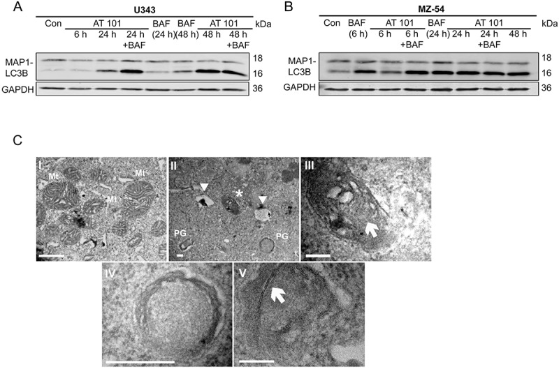
Figure 2.

Effect of AT 101 on the induction of mitophagy in human glioma cells. (A, B) Immunoblotting analysis of MAP1LC3B expression in human glioma cell lines treated with AT 101 (15 µM), bafilomycin A1 (BAF; 10 nM) or DMSO (Con). Bafilomycin A1 was added 4 h before harvest. The time points in brackets refer to the time point when the sample was harvested. (C) Electron microscopy analyses of U343 glioma cells display mitochondria (Mt) with normal morphology, especially with regular cristae structures under control conditions (I)(scale bar: 10 µm; original magnification 20,500x). Upon AT 101 treatment, one could observe the induction of autophagy indicated by the formation of phagophores (PG), autolysosomes (arrowheads) and the engulfment of mitochondria (asterisk) with altered cristae structure (II)(scale bar: 2 µm; original magnification 20,500x). (III)(scale bar: 2 µm; original magnification 60,000x) and (IV)(scale bar: 4 µm; original magnification 105,000x) represent higher magnifications of (II): arrow in (III) points to a mitochondrion that is surrounded by a double membrane thereby indicating its engulfment as a sign of mitophagy; (IV) represents the lower right phagophore in (II). (V)(scale bar: 2 µm; original magnification 160,000x): initiation membranes (arrow) of phagophores are depicted.
