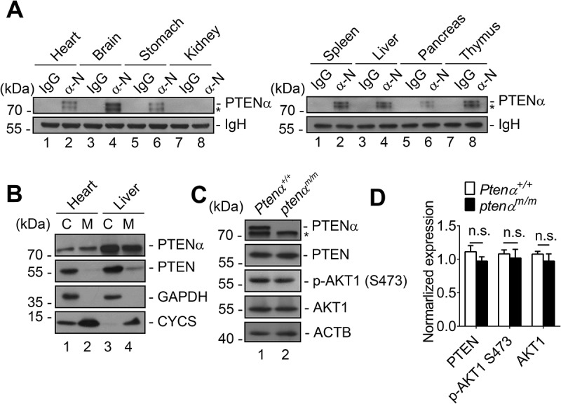
Figure 1.

Confirmation of PTENα deletion in mouse heart. (a) Tissue distribution of PTENα. Analysis of PTENα by immunoprecipitation with PTENα N- terminus-specific antibody (α-N) in various tissue homogenates from 2-months-old wild-type C57BL/6 mice. *, another isoform of PTEN. (b) Subcellular localization of PTENα in mouse liver and heart. C, cytosolic fraction; M, mitochondrial fraction. GAPDH is a cytosolic marker; CYCS is a mitochondrial marker. (c-d) Immunoblot analysis of PTENα, PTEN, AKT1 and p-AKT1 Ser473 in cardiac homogenates. Left ventricles were dissected from 3-months-old mice, and homogenates were immunoblotted with the indicated antibodies. ACTB was used as a loading control. Expression of PTEN, AKT1, and p-AKT1 Ser473 was quantified with ImageJ (n = 3). Data are presented as mean ± SEM; n.s., not significant, p > 0.05 (two-tailed paired Student’s t-test).
