Figure 1.
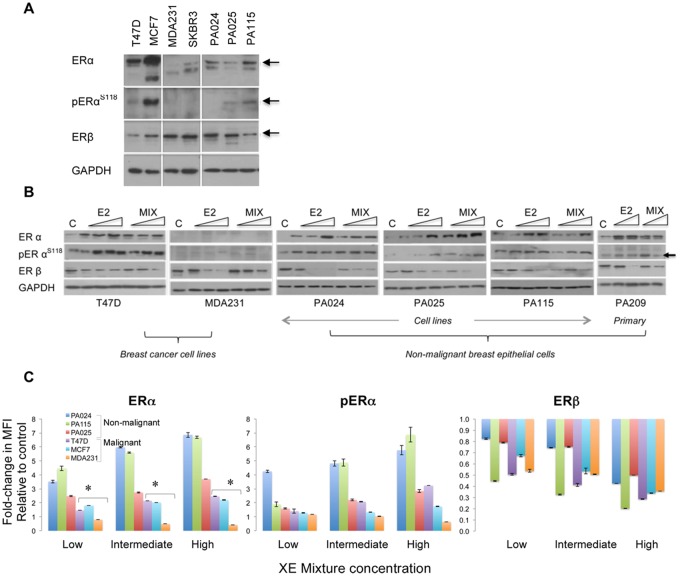
Relative range of cellular perturbations in malignant and nonmalignant breast epithelial cells treated with a mixture of 3 high-consumer use chemicals. A, Baseline levels of ERα and ERβ isoforms shown as representative western blots. Commonly used ERα-positive and ERα-negative breast cancer cell lines are included as controls to compare relative expression levels of each isoform. pERα is designated as pERαS118. GAPDH is the protein loading control accompanying the sample run. When compared with ERα overexpression in ER-positive breast cancer cell lines, HRBECs maintain moderate to low receptor levels. In panels (A) and (B), each subpanel without vertical separation lines represents adjacent samples processed in parallel within the same gel/blot. Horizontal lines within lanes represent discontinuous areas probed with primary antibodies to indicated proteins. B, Concentration-dependent modulation of ER isoform levels in protein lysates of cells treated with 3 increasing concentrations of the test mixture or E2. The primary culture, PA209 was treated with the 2 higher concentrations. Untreated control is denoted as C. Note concentration-dependent induction of ERα in all ER-positive cells but not in ER-negative MDA231 cells. Concurrently, ERβ is reduced by E2 as well as XE mixture treatment in all test cells. C, Quantitation of ERα, pERαS118, and ERβ by multiplexed FACS measurements of fixed whole cells exposed to increasing XE mixture concentration - low (1 nM BPA +1 nM PFOA +10 nM MP), intermediate (10 nM BPA +10 nM PFOA +100 nM MP), and high (100 nM BPA +100 nM PFOA +1 µM MP). Each data point represents 2–4 replicates. As denoted by asterisks, ERα induction in nonmalignant HRBECs (PA024, PA025, PA115) compared with breast cancer cell lines is statistically significant (t test—p ≤ .05). pERαS118 levels in 2/3 mixture-treated HRBEC lines are significantly higher than others. Reduction of ERβ at low mixture concentration relative to untreated control is more pronounced in malignant cells.

